
The Retina Glossary is a guide explaining complex retinal health terms. It helps patients understand conditions, diagnoses, and treatments clearly. Covering retina structure, common conditions, and key terminology, it enhances understanding of eye care.
Overview of the Retina and Its Importance
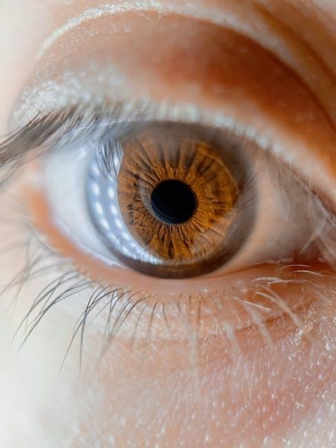
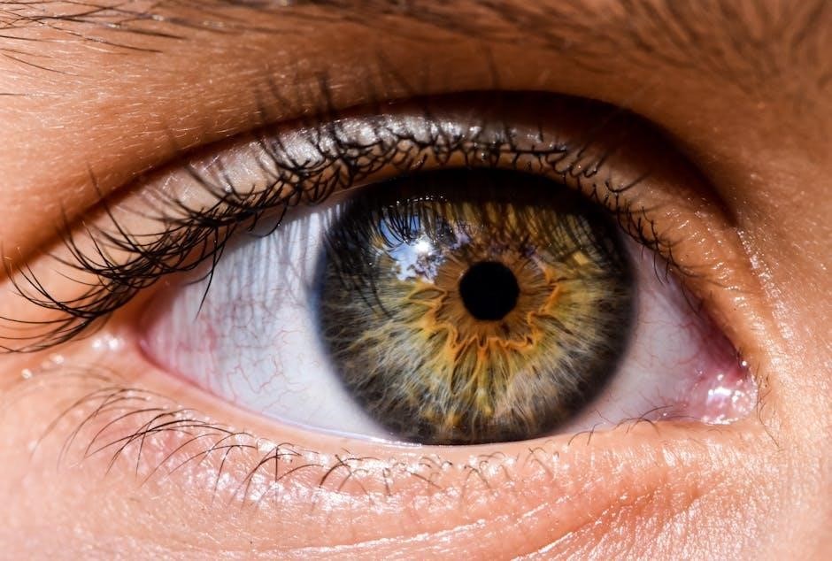
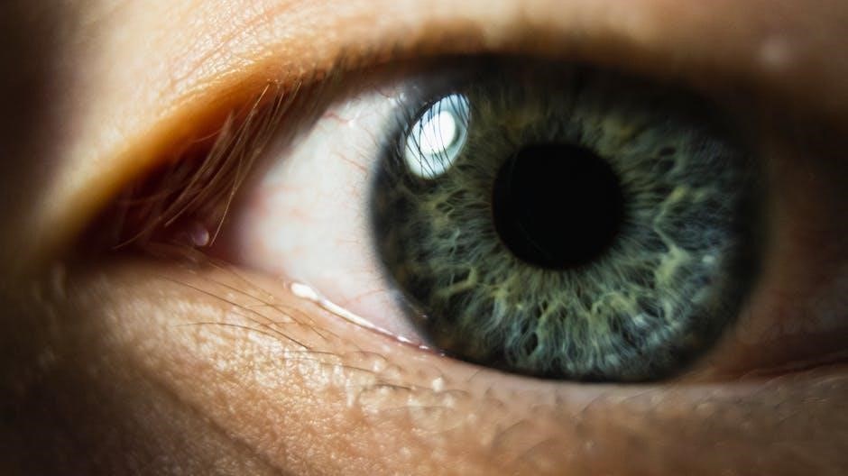
The retina is a vital layer of tissue at the back of the eye, acting as a bridge between light and vision. It contains specialized cells that convert light into electrical signals, which are then sent to the brain via the optic nerve. This process enables us to perceive and interpret visual information. The retina is essential for tasks such as reading, driving, and recognizing faces, making it a critical component of vision health.
Understanding the retina’s function and health is crucial for maintaining clear vision. Conditions like diabetic macular edema and age-related macular degeneration highlight the importance of monitoring retinal health. Protecting the retina through regular eye exams and managing underlying health conditions can prevent vision loss and ensure long-term eye wellness.
Key Terms to Understand Retinal Health
Understanding key terms related to retinal health is essential for grasping common conditions and treatments. Terms like macula, optic nerve, and posterior vitreous detachment (PVD) are fundamental. The macula is the retina’s central part, responsible for sharp vision. The optic nerve carries visual signals to the brain, while PVD refers to the vitreous gel separating from the retina, often a normal aging process. Other terms include myopia (nearsightedness), uveitis (inflammation of the uvea), and retinal ischemia (reduced blood flow to the retina). These terms help patients and professionals communicate effectively about retinal conditions, ensuring accurate diagnoses and appropriate care.
Retina Basics
The retina is a light-sensitive tissue at the back of the eye, converting light into signals sent to the brain. Understanding its structure and function is crucial for eye health.
Definition and Function of the Retina
The retina is a light-sensitive tissue located at the back of the eye, playing a critical role in vision. It consists of specialized cells called photoreceptors (rods and cones) that detect light and convert it into electrical signals. These signals are transmitted to the optic nerve and eventually to the brain, where they are interpreted to form images. The retina is essential for tasks such as reading, recognizing faces, and navigating environments. Its complex structure allows it to process visual information efficiently, enabling us to perceive color, detail, and movement. Understanding the retina’s function is vital for maintaining eye health and addressing conditions that may impair its ability to process light effectively.
Structure of the Retina and Its Layers
The retina is composed of multiple layers, each serving specific functions in visual processing. The outermost layer is the retinal pigment epithelium (RPE), which supports photoreceptor cells by recycling rhodopsin and removing waste products. Beneath the RPE lies the photoreceptor layer, containing rods and cones that detect light and color. The outer nuclear layer houses the cell bodies of these photoreceptors. The inner nuclear layer includes bipolar, Müller, and amacrine cells, which process and transmit signals. The ganglion cell layer contains the axons of ganglion cells, forming the optic nerve. Finally, the inner limiting membrane separates the retina from the vitreous humor. Together, these layers work seamlessly to convert light into electrical signals, enabling vision and visual perception.
Common Retinal Conditions
Common retinal conditions include Diabetic Macular Edema (DME), Age-Related Macular Degeneration (AMD), and Retinal Ischemia. These conditions affect vision by damaging the retina’s structure and function, leading to vision loss if untreated.
Diabetic Macular Edema (DME)
Diabetic Macular Edema (DME) is a condition caused by fluid accumulation in the macula, the central part of the retina, due to diabetic retinopathy. It occurs when high blood sugar levels damage retinal blood vessels, leading to leakage. Symptoms include blurred vision, distorted central vision, and difficulty with tasks like reading or driving. Risk factors include prolonged diabetes, poor blood sugar control, and hypertension. If left untreated, DME can cause irreversible vision loss. Treatment options include anti-VEGF injections to reduce swelling, laser therapy to seal leaking vessels, and corticosteroids. Managing blood sugar levels and hypertension is crucial to prevent progression. Early detection through regular eye exams is vital for preserving vision and quality of life.
Age-Related Macular Degeneration (AMD)
Age-Related Macular Degeneration (AMD) is a progressive eye condition affecting the macula, the central part of the retina responsible for sharp, detailed vision. It is a leading cause of vision loss among people over 50. AMD occurs when the macula deteriorates, disrupting central vision needed for tasks like reading and driving. There are two forms: non-neovascular (dry AMD), characterized by deposits under the retina, and neovascular (wet AMD), marked by abnormal blood vessel growth. Symptoms include blurry vision, distorted lines, and blind spots. Risk factors include age, smoking, and genetics. While there is no cure, treatments like anti-VEGF injections for wet AMD and lifestyle changes can slow progression. Early detection through regular eye exams is critical for managing AMD and preserving vision.
Retinal Ischemia and Its Implications
Retinal ischemia occurs when blood flow to the retina is obstructed, leading to oxygen deprivation and tissue damage. This condition can result from blockages in retinal arteries or veins, often due to blood clots, hypertension, or diabetes. Symptoms include sudden vision loss, blind spots, or blurry vision. If untreated, retinal ischemia can cause permanent damage, leading to severe vision impairment. It is a medical emergency requiring immediate attention. Treatments may involve addressing the underlying cause, such as managing blood pressure or diabetes, or procedures like intraocular injections to restore blood flow. Early diagnosis is critical to prevent long-term vision loss, emphasizing the importance of regular eye exams, especially for high-risk individuals. Retinal ischemia underscores the delicate balance of retinal health and the need for prompt intervention.

Retinal Terminology
Retinal terminology refers to the language used to describe the retina’s structures, functions, and conditions. It includes terms like macula, optic nerve, and photoreceptors, aiding in understanding retinal health and diseases. Understanding this vocabulary helps patients and professionals communicate effectively about diagnosis, treatment, and management of retinal conditions.
Optic Nerve and Its Role

The optic nerve is a vital component of the visual system, serving as the pathway for visual information from the retina to the brain. Comprising over a million nerve fibers, it transmits signals that enable us to interpret visual stimuli. The optic nerve exits the eye through the optic disc and is essential for relaying images to the brain for processing. Damage to this nerve can lead to vision impairment or blindness. Conditions like glaucoma, characterized by increased intraocular pressure, can adversely affect the optic nerve, highlighting its crucial role in maintaining eye health. Understanding the optic nerve’s function is fundamental for appreciating how visual information is processed and the importance of protecting this critical structure.

Macula: The Center of the Retina
The macula is a small, highly sensitive area at the center of the retina responsible for central vision, fine detail, and color perception. It enables activities like reading, driving, and recognizing faces. The macula contains a high concentration of photoreceptor cells, particularly cones, which are crucial for sharp, detailed vision. Any damage to this area can lead to significant visual impairment, such as blurred or distorted central vision. Conditions like age-related macular degeneration (AMD) specifically affect the macula, highlighting its importance in maintaining visual clarity and quality. Understanding the macula’s role is essential for appreciating how the retina processes visual information and the impact of its dysfunction on daily life.
Posterior Vitreous Detachment (PVD)
Posterior vitreous detachment (PVD) is a common age-related condition where the vitreous gel separates from the retina. The vitreous is a clear gel that fills the center of the eye, and as we age, it naturally shrinks and pulls away from the retina. While PVD itself is typically harmless, it can sometimes lead to symptoms like flashes of light or an increase in floaters. In some cases, it may increase the risk of retinal tears or detachment. PVD is more common in individuals over 50 and those who are nearsighted. It is important to monitor symptoms and consult an eye care professional if they suddenly worsen, as early detection of related complications can prevent severe vision loss.
Eye-Related Terms
Eye-related terms describe conditions and structures vital to understanding eye health, such as myopia, uveitis, and anterior chamber angle, each impacting vision uniquely and significantly.
Myopia: Causes and Effects
Myopia, or nearsightedness, is a common eye condition where distant objects appear blurry due to the eyeball being too long or the cornea too curved. Causes include genetics, prolonged near work, and insufficient outdoor activity. Environmental factors, such as excessive screen time, also contribute. The condition often develops in childhood and progresses until adulthood. Effects of myopia extend beyond vision impairment; severe cases increase the risk of retinal detachment, glaucoma, and cataracts. Early diagnosis and corrective measures, like glasses or contact lenses, are essential to manage symptoms and prevent complications. Understanding myopia is crucial for maintaining eye health and addressing its broader implications.
Uveitis: Inflammation of the Uvea
Uveitis is the inflammation of the uvea, the eye’s middle layer, which includes the iris, choroid, and ciliary body. This condition can cause eye pain, redness, sensitivity to light, and blurred vision. It is classified into anterior, intermediate, and posterior uveitis, depending on the affected part. Causes include infections, autoimmune disorders, or trauma. If untreated, uveitis can lead to complications like glaucoma or vision loss. Treatment typically involves anti-inflammatory medications, such as corticosteroids, and addressing underlying causes. Prompt medical attention is crucial to manage symptoms and prevent long-term damage. Uveitis is a significant condition in retinal health, as it can impact the retina and overall vision quality.

Anterior Chamber Angle
The anterior chamber angle is the space between the iris and the cornea at the front of the eye. It is a critical area for aqueous humor drainage, which helps maintain intraocular pressure. The angle is formed by the junction of the iris root and the cornea. A narrow or closed anterior chamber angle can lead to increased intraocular pressure, potentially causing conditions like angle-closure glaucoma. An open angle allows proper drainage, preventing such complications; Eye exams often assess the angle to ensure healthy aqueous flow and overall eye function; Understanding this structure is vital for diagnosing and managing glaucoma and other eye disorders.

Treatments and Procedures
Common treatments include laser therapy, anti-VEGF injections, and vitrectomy surgery. Early detection and personalized care are crucial for managing retinal conditions effectively.
Laser Therapy in Retinal Care
Laser therapy is a cornerstone in managing retinal conditions, offering precise and minimally invasive treatment. It works by targeting damaged or abnormal blood vessels, reducing leakage and inflammation. Conditions like diabetic retinopathy and macular edema benefit significantly from this approach. The procedure involves focusing a high-intensity light beam to seal blood vessels, prevent further damage, and promote healing. Different types of lasers, such as argon and diode, are used depending on the condition. Laser therapy is favored for its localized impact, minimizing harm to surrounding tissue. Regular follow-ups are essential to monitor treatment efficacy. Early intervention with laser therapy can preserve vision and prevent complications, making it a vital tool in modern retinal care.
Role of the Retina Specialist
A retina specialist is an ophthalmologist with advanced training in diagnosing and treating retinal disorders. They play a critical role in managing conditions like macular degeneration, diabetic retinopathy, and retinal detachments. Using tools such as optical coherence tomography (OCT) and fluorescein angiography, they detect early signs of retinal damage. Their expertise includes performing intricate surgeries, such as vitreoretinal procedures, and administering injections for conditions like macular edema. Retina specialists also conduct comprehensive eye exams to monitor disease progression and develop personalized treatment plans. Their goal is to preserve vision, prevent further damage, and improve quality of life for patients. Regular follow-ups with a retina specialist are essential for maintaining retinal health and addressing concerns promptly.
Importance of Understanding Retinal Health
Understanding retinal health is critical for maintaining vision and preventing vision loss. This glossary provides essential knowledge to empower individuals in managing and protecting their eye health effectively.
Understanding retinal health is vital for preserving vision and addressing potential issues early. The retina, being the light-sensitive layer at the back of the eye, plays a crucial role in vision clarity and acuity. Conditions like age-related macular degeneration (AMD) and diabetic macular edema (DME) highlight the need for awareness. By familiarizing oneself with retinal terminology, individuals can better recognize symptoms, seek timely care, and prevent vision loss. This knowledge also fosters improved communication with eye care professionals, enabling more informed decisions. Regular eye exams and lifestyle adjustments, such as managing diabetes and avoiding smoking, are key preventive measures. Educating oneself about retinal health empowers individuals to take proactive steps in safeguarding their vision and overall eye wellness.

Resources for Further Learning
For those seeking to deepen their understanding of retinal health, numerous resources are available. The American Academy of Ophthalmology offers comprehensive guides and articles on retinal conditions. Additionally, the National Eye Institute provides detailed information on retinal diseases and treatments. Books like Clinical Ophthalmology and online courses on platforms like Coursera can also serve as valuable educational tools. Many eye care clinics offer downloadable pamphlets and videos explaining retinal health in simple terms. Staying informed through reputable sources ensures better awareness and proactive care for eye wellness. Regularly consulting with a retina specialist is also encouraged for personalized advice and updates on the latest research.